
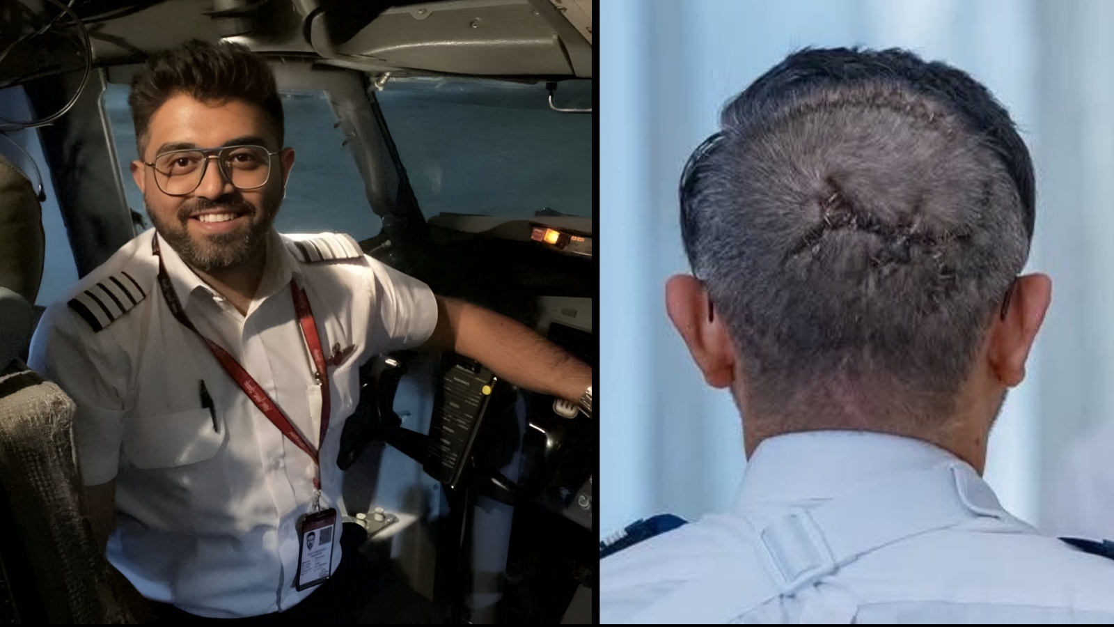
Captain Smit Machchhar, the Indian-origin pilot of Flydubai flight FZ1073, who was allegedly attacked by his co-pilot, has been awarded the First Class Order of Zayed II by the UAE President. Videos from the ceremony have drawn attention to the serious head injury he sustained. A neurosurgeon explains the medical complexities that may have made his injury particularly challenging to treat.
In a post on X, Dr Sanat Dixit described the injury as an “occipital compound depressed skull fracture” treated with a “wide craniotomy”. He also highlighted its proximity to the posterior superior sagittal sinus, a major blood-draining channel in the brain.
But what does this injury actually mean, and why is its location so concerning? We reached out to Dr Ravindra Srivastava, Director and Neurosurgeon at ShardaCare – Healthcity, seeking answers.
DISCLAIMER: This article is based on information from the public domain and/or the experts we spoke to. Always consult your health practitioner before starting any routine.
What is an occipital compound depressed skull fracture?
As a neurosurgeon, let me tell you how frightening this injury actually is – an occipital compound depressed skull fracture treated with a WIDE craniotomy (outer staple line).
What’s scary isn’t the fracture; it’s what’s adjacent to the damaged skull. The axe blows (that tree-branch scar at the center) hit right over the posterior superior sagittal sinus, a large vein that runs lengthwise between the 2 cerebral hemispheres and carries blood from the brain to the heart.
Penetrating injuries to the sinus can carry upwards of a 40% morbidity/mortality – partially because a transection of the sinus can be difficult to repair and can result in extreme blood loss, venous air embolism, or thrombosis – all of which can be fatal.
There’s scary, and then there’s the “you go to elevate the fracture and risk unroofing the sinus, losing 2 liters of blood in a minute” scary.
The surgeons likely had their hands full and did an amazing job. Divine providence called many heroes into service that day. Damn fine work.
धन्यवाद
— Sanat Dixit MD FACS (@sdixitmd) October 28, 2026
“An occipital compound depressed skull fracture occurs when the bone at the back of the skull breaks, causing fragments to move inward, along with an associated open wound,” Dr Srivastava tells indianexpress.com.
In simple terms, this is more than a crack in the skull. The broken bone fragments are pushed inward, towards the brain, while the associated open wound can expose the underlying structures to further damage.
“These fragments can damage the underlying brain tissue, its protective coverings (meninges) and nearby blood vessels,” the expert adds.
The meninges are the protective membranes surrounding the brain. Depending on the extent of the injury, damage to these structures can lead to bleeding, brain swelling, and infection.
Story continues below this ad
“It is a serious condition requiring urgent neurosurgical assessment and, in many cases, surgical intervention to prevent further complications.”
Why is the location of the fracture particularly dangerous?
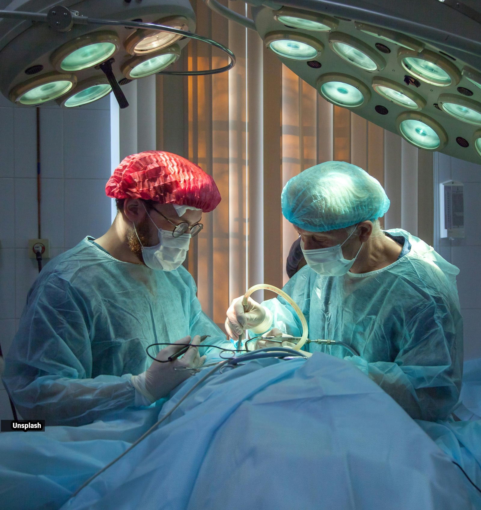 Surgeons must carefully manage the depressed bone fragments without damaging this major venous channel (Image: Unsplash)
A major concern highlighted by Dr Dixit was the injury’s proximity to the posterior superior sagittal sinus, an important structure that helps drain blood from the brain.
“The superior sagittal sinus is a major venous channel running along the midline of the brain, responsible for draining blood from the brain towards the heart,” Dr Srivastava explains.
Damage to this channel can have serious consequences, “potentially causing significant blood loss, air embolism or thrombosis.”
Story continues below this ad
Air embolism occurs when air enters the bloodstream and obstructs blood flow, while thrombosis refers to the formation of a blood clot. Both can obstruct normal circulation. Damage to the sinus can also interfere with the drainage of blood from the brain.
“It can also disrupt normal venous drainage, resulting in brain swelling and increased intracranial pressure,” Dr Srivastava further notes. (Intracranial pressure, here, refers to the pressure inside the skull, which can rise when the brain swells.)
This proximity also makes surgery particularly challenging. Surgeons have to carefully handle the “depressed bone fragments” without damaging the sinus while trying to reach and treat the injured area.
“Any injury to it can cause rapid blood loss and complications such as air embolism, thrombosis and impaired venous drainage,” he added. This is why careful surgical planning, controlled removal of the fractured bone fragments and preparedness to manage blood vessel injuries are essential.
Story continues below this ad
What does a wide craniotomy involve?
Dr Dixit also described the injury as having been treated with a wide craniotomy. “A wide craniotomy involves temporarily removing a larger section of the skull to provide better access and visibility of the injured area,” Dr Srivastava explains.
This allows surgeons to reach the affected area and assess the damage beneath the skull. “In this case, it may have been performed to manage the fracture, remove displaced bone fragments, control bleeding and assess or repair damage to the underlying structures,” he further explains.
However, surgery does not necessarily mean that all risks are over. “Potential complications include infection, seizures, bleeding and neurological deficits,” Dr Srivastava points out.
The patient’s long-term recovery depends largely on the extent of the initial brain injury and how they respond to treatment.
Story continues below this ad
Dr Srivastava acknowledges that this was a “particularly complex injury” because of its proximity to a major venous drainage channel.
“The priority in such cases is to protect the underlying brain while preserving normal blood circulation. Timely intervention, careful surgical management and close postoperative monitoring are crucial for recovery,” Dr Srivastava lastly notes.
DISCLAIMER: This article is based on information from the public domain and/or the experts we spoke to. Always consult your health practitioner before starting any routine.

Leave a Reply